Gastroenterology Research, ISSN 1918-2805 print, 1918-2813 online, Open Access
Article copyright, the authors; Journal compilation copyright, Gastroenterol Res and Elmer Press Inc
Journal website http://www.gastrores.org

Original Article

Volume 11, Number 4, August 2018, pages 274-279


Feasibility of Underwater Endoscopic Mucosal Resection for Colorectal Lesions: A Single Center Study in Japan

Figures

Figure 1.
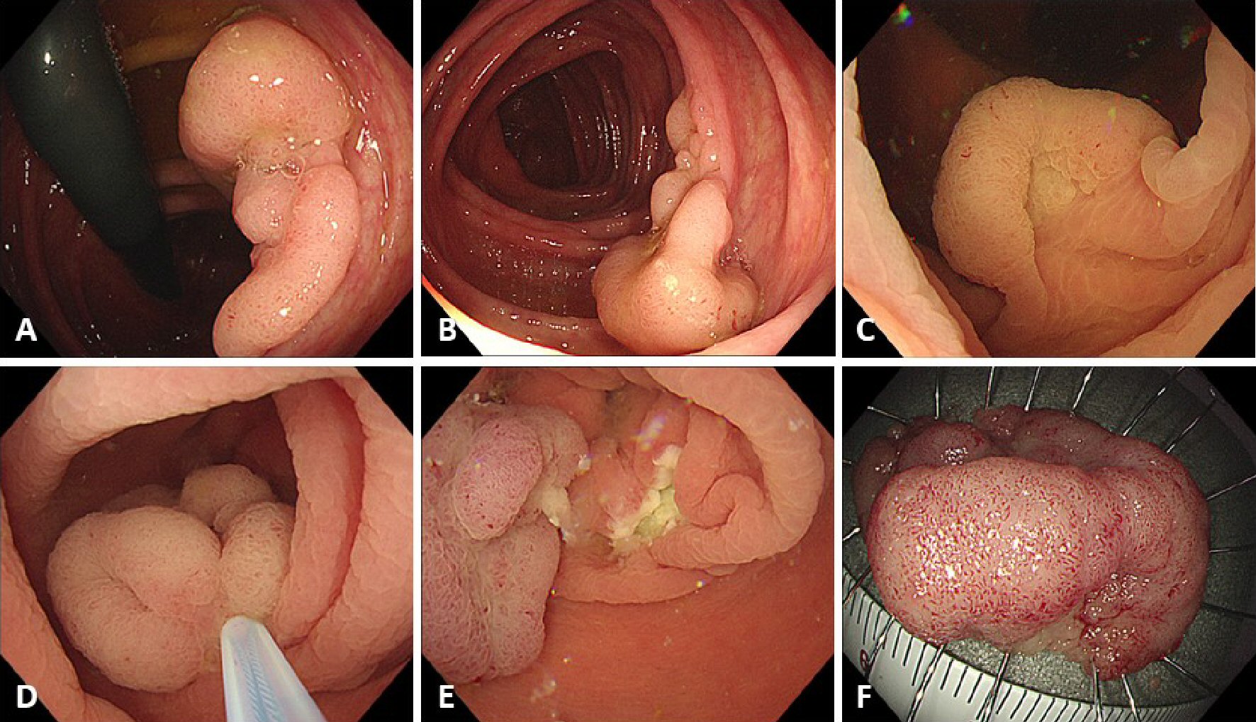
Figure 1. Procedure for underwater endoscopic mucosal resection (U-EMR). (A, B) A sessile lesion about 30 mm located in the ascending colon. (C) The lumen at the site of the lesion was filled with water, using the water-jet system, and the lesion was floated in the lumen. (D) The lesion was snared, using a 33-mm snare, and the lesion was tightened-up. (E) The mucosal defect after U-EMR was usually smaller than after conventional EMR. (F) Resected specimen after successful en bloc resection.
Figure 2.
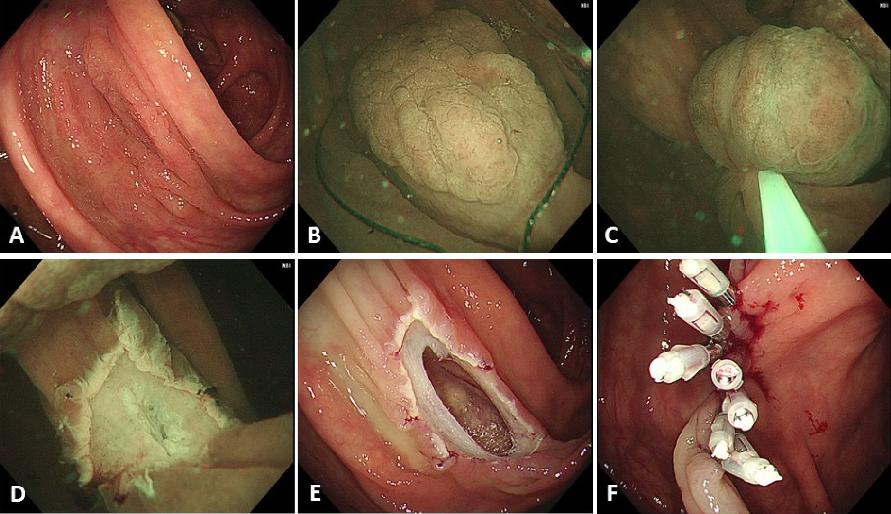
Figure 2. A case of perforation. (A) A laterally spreading, granular type lesion about 30 mm located in the ascending colon. (B) Adequate floating of the lesion could not be achieved and, therefore, submucosal injection, using normal saline, was performed before snaring. (C) The lesion was snared, using a 33-mm snare, and the lesion was tightened-up. (D) En bloc resection was achieved but the muscularis propria was slightly injured at the center of the mucosal defect. (E) An obvious perforation was identified after gas insufflation in the lumen. (F) Endoscopic clipping was performed, and surgical intervention was not required.

Tables

Table 1. Patient and Lesion Characteristics
 
Number of cases64 lesions in 38 cases
Mean age, years (range)68.6 (25 - 90)
Male sex, N (%)26 (68)
Mean size of lesions, mm (range)16.2 (6 - 40)
  < 10 mm, N (%)8 (13)
  10 - 19 mm, N (%)34 (53)
  20 - 29 mm, N (%)15 (23)
  ≥ 30 mm, N (%)7 (11)
Location
  Cecum, N (%)9 (14)
  Ascending colon, N (%)12 (19)
  Transverse colon, N (%)12 (19)
  Descending colon, N (%)7 (11)
  Sigmoid colon, N (%)17 (27)
  Rectum, N (%)7 (11)
Morphology
  0-Is, N (%)33 (52)
  0-Ip, N (%)10 (16)
  0-IIa, N (%)18 (28)
  0-IIa+Is, N (%)3 (5)
Histology
  Low-grade adenoma, N (%)31 (48)
  High-grade adenoma, N (%)8 (13)
  Mucosal cancer, N (%)11 (17)
  Submucosal cancer, N (%)4 (6)
  Sessile serrated adenoma/polyp, N (%)4 (6)
  Traditional serrated adenoma, N (%)1 (2)
  Hyperplastic polyp, N (%)3 (5)
  Inflammatory polyp, N (%)2 (3)


Table 2. Study Outcomes
 
‡Complete resection rate was defined by a negative pathological margin of resected specimen that was obtained by successful en bloc resection.
En bloc resection rate
  Overall, %81 (52/64 lesions)
  < 10 mm, %100 (8/8)
  10 - 19 mm, %94 (32/34)
  20 - 29 mm, %53 (8/15)
  ≥ 30 mm, %57 (4/7)
Complete resection rate‡
  Overall, %54 (32 of 59 neoplastic epithelial lesions)
  < 10 mm, %88 (7/8)
  10 - 19 mm, %63 (20/32)
  20 - 29 mm, %25 (3/12)
  ≥ 30 mm, %29 (2/7)
Post procedural bleeding, N (%)3 (5)
Perforation, N (%)1 (2)


Table 3. Cases of Submucosal Cancer in This Study
 
LST-NG: laterally spreading tumor, non-granular type; LST-G: laterally spreading tumor, granular type; N/A: not applicable.
Age (years)67719085
SexFMMM
LocationAscending colonAscending colonSigmoid colonCecum
Size (mm)40301215
Morphology0-IIa+Is (LST-G)0-Is0-IIa (LST-NG)0-Is
En bloc resectionNoNoYesYes
Submucosal invasion depth (µm)2,5002,8002,000150
Lateral marginN/AN/A+-
Base margin----